



AI supports tumour burden assessment in PET imaging – Atostek implemented a user interface for Turku PET Centre
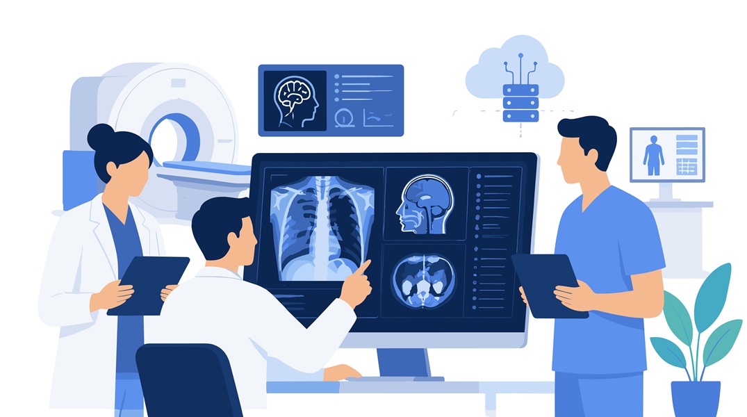
Atostek has implemented a user interface for the Wellbeing Services County of Southwest Finland that brings AI algorithms for cancer imaging into Turku PET Centre’s research work. The solution combines the results of algorithms that identify cancerous changes and organ regions into a single view, where specialists can review and correct the AI-generated findings. The aim is to streamline the processing of imaging data and support tumour burden assessment.
Cancer imaging involves processing large volumes of image data. PET/CT and PET/MRI examinations can be used to detect, for example, cancer metastases, but interpreting the images requires precision and expertise. AI can support this work by automatically identifying findings and calculating metrics that describe the overall picture of the disease.
The Wellbeing Services County of Southwest Finland and Turku PET Centre have developed AI algorithms that identify tumours in PET images. Atostek built a user interface that enables the algorithm results to be reviewed, corrected and utilised in research work.
The user interface combines algorithm results
The user interface developed by Atostek combines the results of the algorithms into a single view. It makes it possible to review in which organs or anatomical regions the AI-identified changes are located and what their volume is.
The user interface is based on OHIF software (Open Health Imaging Foundation), an open-source solution for viewing medical images. Atostek modified and extended the OHIF-based interface to suit this specific use case. AI-assisted software development methods were also used during the development work.
“This has been a highly interesting project in which AI is being harnessed for an important purpose: supporting cancer research. Atostek was responsible for building a user interface that brings the algorithm results to specialists and makes them part of the research workflow. It has been great to work on this together with a skilled team developing something new,” says Juho Leppämäki, Project Manager at Atostek.
A physician can review the results produced by the algorithm in the images and correct them if necessary. This creates a specialist-verified version that can be used for further training of the algorithms.
The aim is to make physicians’ work easier
The aim of the solution is to speed up and facilitate physicians’ work and support tumour burden assessment. Based on the findings, the software calculates, for example, tumour volumes in different organ regions. This supports research in assessing disease spread, treatment response and disease progression.
“Atostek’s technical expertise played a key role in bringing the AI algorithms we have developed into the practical workflow of research. At the same time, this lays the foundation for increasingly accurate AI methods that may in the future support tumour burden assessment and PET imaging analysis in an increasingly systematic way,” says Jukka Kemppainen, Professor of Clinical Physiology and Nuclear Medicine at the Turku University Hospital.
The software will be taken into research use at Turku PET Centre. It is not a solution that replaces the physician’s work, but a tool that supports specialist assessment and research.
The project has involved the Wellbeing Services County of Southwest Finland, Turku PET Centre and Atostek.